Spine Surgery
- Home
- Spine Surgery

ANTERIOR CERVICAL
DISKECTOMY AND FUSION
- Perhaps the most common surgery performed by neurosurgeons.
- By making a small semilunar incision on the neck in the groove or wrinkle, one safely accesses the anterior spine and removes disks, osteophytes, and even whole “vertebral” bodies. A square block (otherwise known as a “cage”) is placed to hold up the space. There is often a plate or a small set of screws/blades that hold this cage in place.
- This surgery has very low risks in general is ~ 85-95% successful. If more than 3 levels of disk removal are needed, one tends to add posterior screws or instrumentation to ensure no movement and stability
- Typically, one level surgery is outpatient or overnight stay. 3 level surgery may require 1-2 days in the hospital as the neck swelling recovers, mainly in speaking and swallowing.
CERVICAL ARTHROPLASTY
THE “CERVICAL ARTIFICIAL DISK”
- This surgery is an excellent option for young patients who have disk osteophytes of disk-related narrowing of canal and nerves (“radiculopathy”).
- Surgery must fulfill two rules: (a) the posterior (“back of neck”) must have good joints and no excessive arthritis or motion; (b) patient’s neck should not have a moderate or severe deformity.
ANTERIOR LUMBAR
INTERBODY AND FUSION
- Typically performed for patients who are having multiple levels of fusion with degeneration, and, who are at risk for pseudoarthrosis (failure of fusion and continued pain), and, who are “flat” in the back and benefit from curvature improvement
- Very helpful technique in revision cases where there is poor fusion and too much scar around the nerves for proper posterior technique for discectomy.
- Surgery often increases the height of the patient
- This surgery is extremely surgeon-dependent and the risks of the surgery could be high in some hospitals and very low in others. My vascular surgeon colleagues and I have performed > 500 of such operations over almost 20 years with high safety. However, these surgeries and patients are carefully chosen
POSTERIOR LUMBAR
INTERBODY AND FUSION
- Typically performed for patients who are having one or multiple levels of fusion with degeneration, and, who are at risk for pseudoarthrosis or continued instability or motion (failure of fusion and continued pain or slippage of disks), and, who do not have curvature issues or scars that prevent me from working around the nerves.
- An expandable cage is placed after the discectomy that works as a “lifter” of the disk space to acquire height.
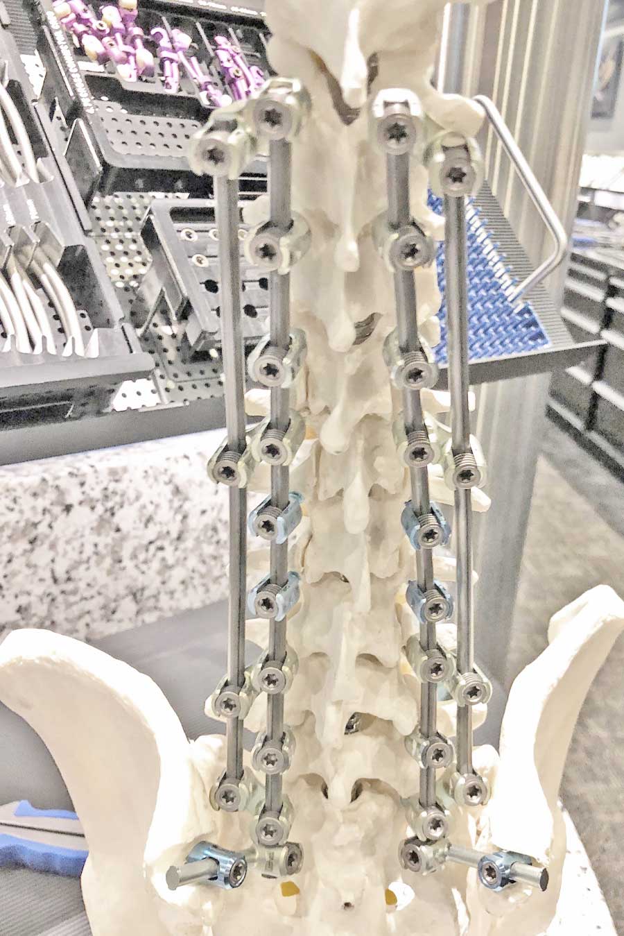
Dual Rod construct for trauma or tumor repair

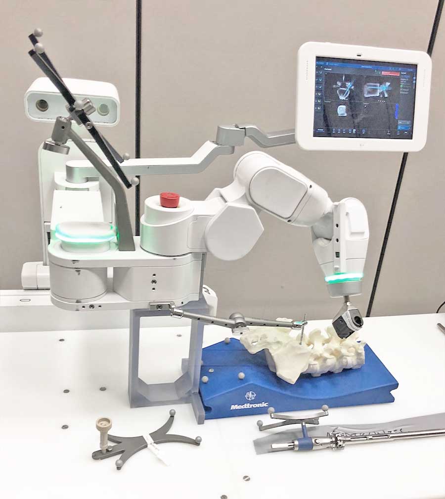
Excelsius and Mazor X

LUMBAR ARTHROPLASTY
“THE LUMBAR ARTIFICIAL DISK”
- Currently, due to lack of a reliable FDA-approved system, I am not performing this operation. Current artificial disks on the market have a relatively high complication rate that I am currently avoiding
SPINAL
CORPECTOMY
- This is a removal of the bone and the disks above and below the bone of an entire level of the spine. The standard approach to a cervical or lumbar corpectomy is similar to that of anterior cervical discectomy or anterior lumbar discectomy.
- Thoracic corpectomies are typically performed either from the posterior approach or from the anterior (through the lung and chest) and posterior approach. I tend to avoid the lung or chest and perform the removal of the bone or cancer or tumor from the posterior approach alone through a highly specialized corridor called the Transpedicular Approach.
- In certain cancers, it is crucial to remove the entire bone as a block and a ‘spondylectomy’ is used.
SPINAL SURGURY
MICRODISKECTOMY
- typically done using a small tube and a half-inch incision, this surgery is common and leaves little scar. This surgery is often referred to as the “laser incision” or the “minimally invasive” surgery.
- There is a separate method to remove disks with no muscle dissection utilizing an endoscope that comes at ~ 40 deg to the spine. This method has been popularized by a company called Joimax. I am currently working on a robotic-guided method as well.
LAMINOTOMY, LAMINECTOMY
POSTEROLATERAL OSTEOTOMY
- These terms describe the amount of posterior bone that is removed to free nerve and spinal cord structures. Sometimes, a tiny ½ inch hole is all that is needed. In other cases – such as scoliosis – the bone, ligaments and the entire posterior joint (the “facet”) need to be disrupted to achieve a curve correction.
PEDICLE SCREWS
INSTRUMENTATION
- In 2003, Dr. Fritzell and the Swedish Lumbar Spine Study Group published the results of 211 patients aged 25-65 who underwent spine surgery for the same condition but in 3 separate manners; one group had just a laminectomy, another a laminectomy with bone growth overlay, and a third group with a laminectomy and then pedicle screws instrumentation to promote the bone growth further. The study demonstrated a higher success rate in the instrumentation and fusion group AS LONG AS there were no hardware placement or associated complications. This study showed that, as long as instrumentation is used properly and accurately, good outcomes could be seen.
